Material Resin
Quantity 1 pcs
Price Range $1-100
Lead Time 2 workdays
Gallery
About Project
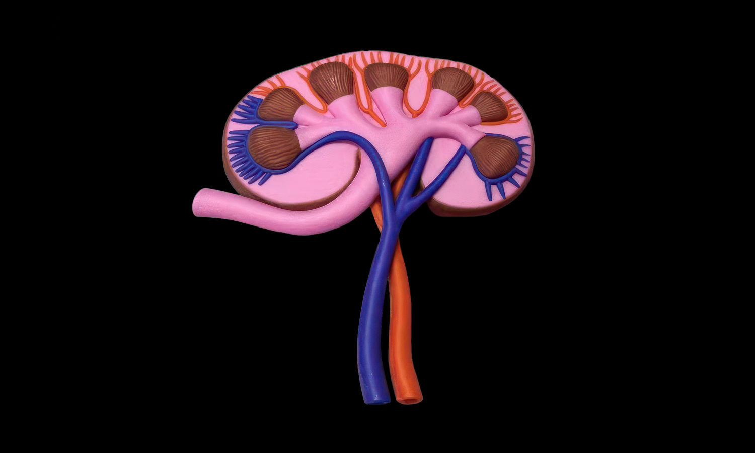
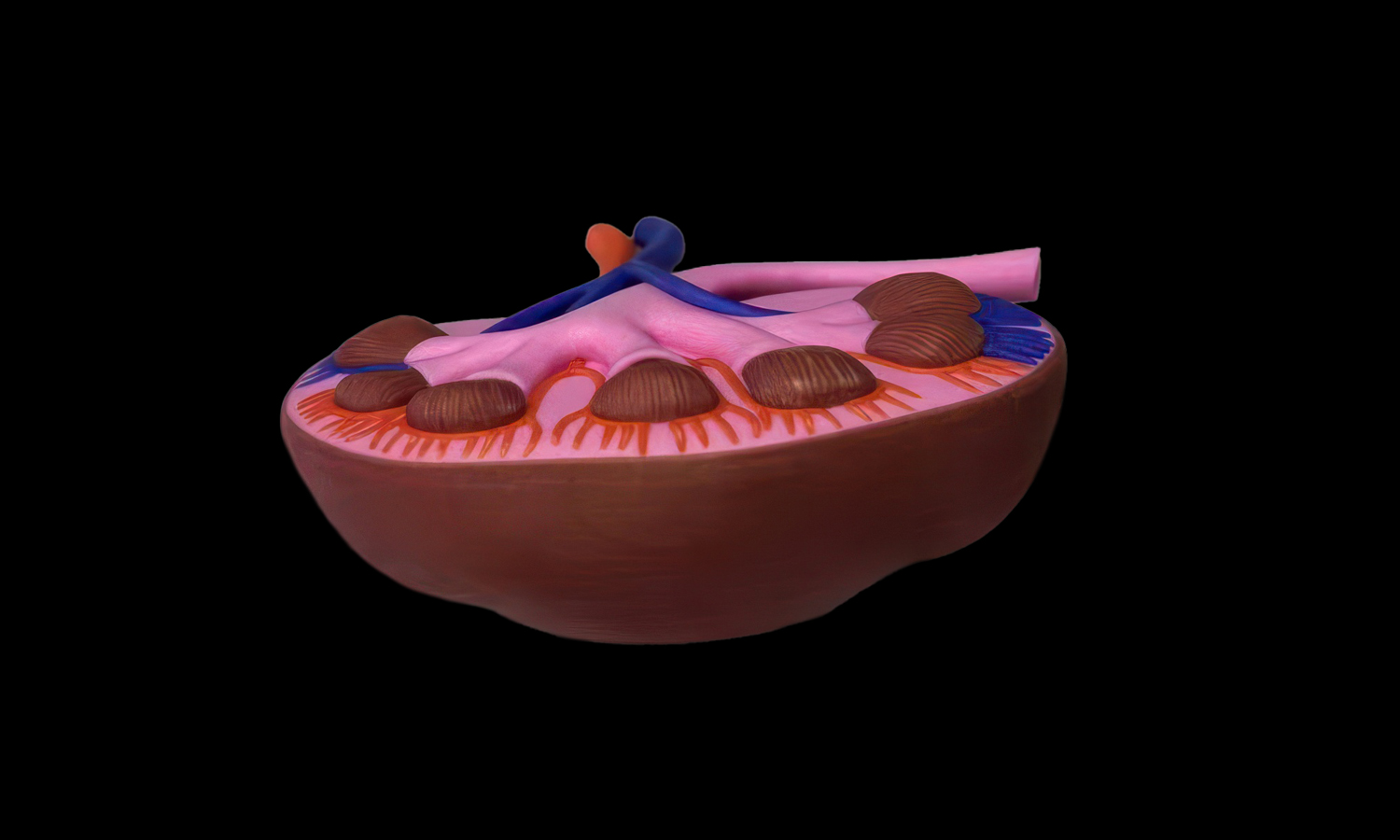
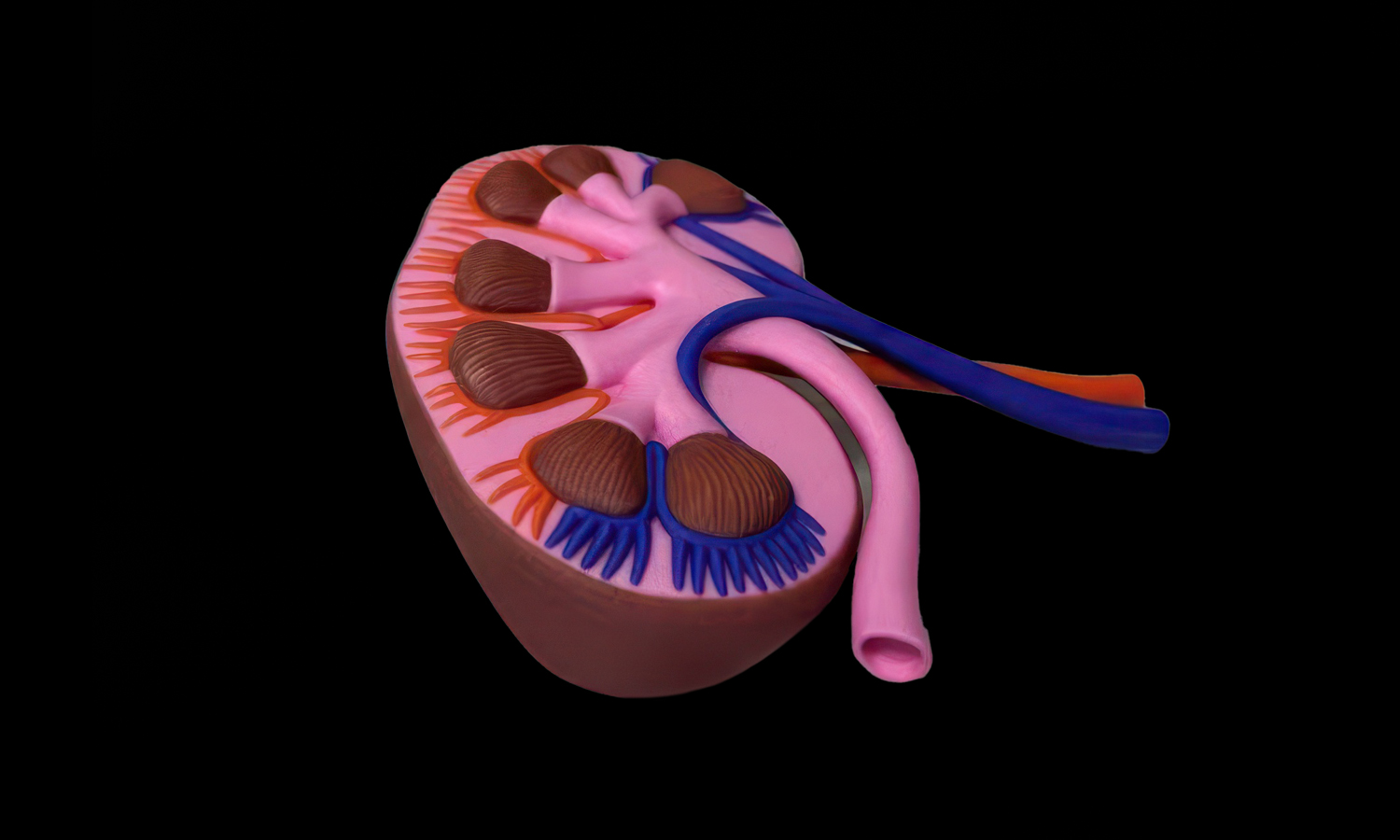
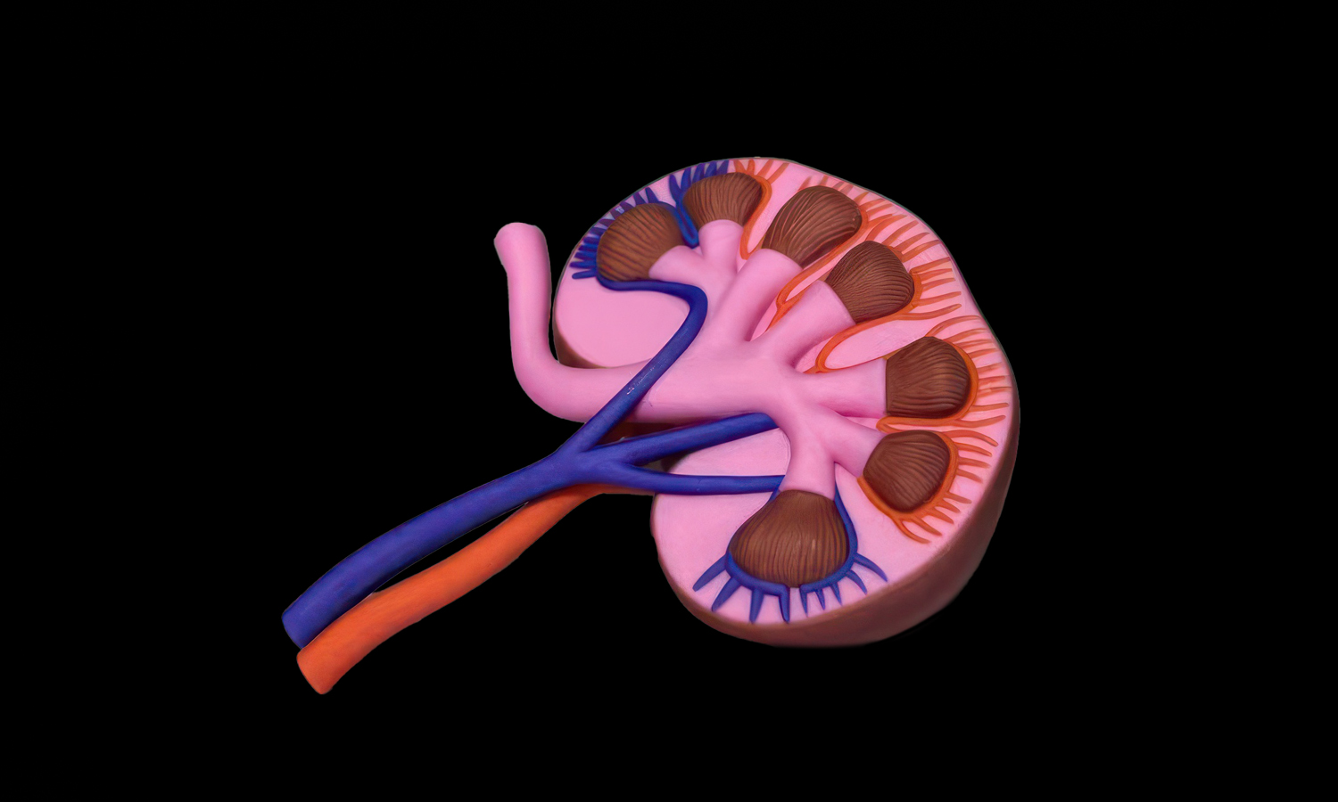
Anatomy models are most useful when they make complex structures easier to see at a glance. This full-color kidney anatomy model presents the organ in a clean sectional view, showing the outer kidney shape, internal tissue regions, renal vessels, ureter, and branching collecting structures with clear color separation.
The model uses pink, brown, red, blue, and purple color zones to help different anatomical parts stand out from each other. Compared with a flat diagram, a physical model gives students, educators, clinicians, and visitors a more direct way to understand spatial relationships inside the kidney, especially the paths of the vessels and the layered internal structure.
Mimaki full-color 3D printing was a strong fit for this project because it can reproduce color-coded anatomical regions, fine vessel branches, raised tissue textures, smooth curved surfaces, and clean educational details directly from the digital model. For medical educators, research teams, museums, and healthcare brands developing anatomical teaching models, FacFox provides professional full-color 3D printing services for medical visualization and display prototypes. You can get an instant quote online or email us to discuss your projects.
Solution
- Step 1: The kidney anatomy model was reviewed before printing, and areas such as the renal section, vessel branches, ureter, tissue textures, and color-coded anatomical regions were checked for clarity and manufacturability.
- Step 2: Full-color material settings were assigned to different anatomical areas, including the pink internal tissue, brown renal pyramids, red and blue vessels, purple branches, and outer kidney wall.
- Step 3: Support structures were generated around the raised vessels, ureter, internal branches, and curved sectional surface to keep the delicate anatomical features stable during printing.
- Step 4: The model was produced with Mimaki full-color 3D printing, where UV-cured photopolymer materials were printed layer by layer to form the color-coded anatomy and surface details in one integrated part.
- Step 5: After printing, support material was carefully removed from the vessel branches, ureter, inner grooves, and raised tissue textures without damaging the fine educational details.
- Step 6: The finished kidney model was cleaned and inspected from multiple angles to confirm that the color separation, anatomical structure, vessel paths, and sectional details were clearly reproduced.