Researchers from Nottingham Trent University (NTU) have developed realistic 3D printed heart and lung models that can bleed, beat and breathe like their real counterparts.
Designed for organ transplant training, the lifelike models reportedly reflect the tactile qualities of a human heart and can be produced with various tissue hardness levels. Using the models, medical professionals can plan surgeries and safely research and teach transplant procedures, without the risk of complications.
The project, which was led by research fellow Richard Arm, leveraged 3D scans of both healthy and diseased human hearts to 3D print the models to a high level of accuracy.
“The aim is to give surgeons the opportunity to learn the technical aspects of organ transplant surgery and experience the tactile aspects of removing a failing heart and connecting a different healthy one, identifying and suturing the vessels that keep the donor heart in place,” explained Arm.
“This technology can simulate bleeding like a normal heart to provide the actual experience and limited visibility that surgeons must face on the operating table.”
The project was funded by the Freeman Heart and Lung Transplant Association (FHLTA) and was presented at the annual meeting of the Society for Cardiothoracic Surgery last month.

 Senior research fellow Richard Arm holding a 3D printed heart model. Photo via Nottingham Trent University.
3D printing realistic organ models
Currently, most trainee surgeons practice with cadavers and animal organs, as existing models aren’t realistic enough. The NTU researchers claim that their models are the first to offer the realism required for training purposes.
To ensure high levels of realism, CT data is gathered from real patients, which is then used to 3D print the physical models.
While researchers at the NTU have developed other fake organs in the past, Arm believes this to be their most advanced yet, representing the “first example of the next generation in synthetic organs where realistic internal detailing crucially includes all the important, hidden blood vessels and heart valves.”
“There’s simply nothing like this in the world right now, so we’re currently working alongside British surgeons and global retailers to trial this next generation of medical models,” stated Arm. “It is really important because heart transplantation is a specialist skill.”
The organ models feature bleeding vessels and the pericardium, a thin fluid-filled sac that surrounds the heart. This allows trainee surgeons to accurately simulate the process of stemming blood flow, which is required before making incisions and removing diseased organs. Additionally, the team’s lung models have been designed to mimic natural breathing patterns.

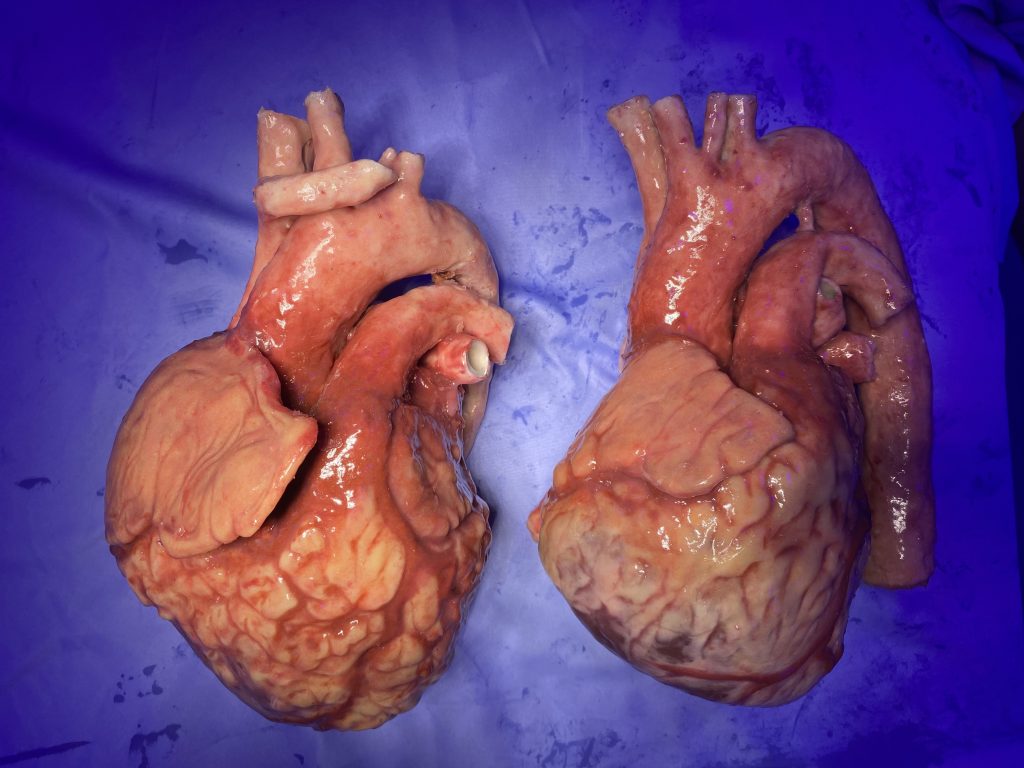 NTU 3D printed heart models. Photo via Nottingham Trent University.
It is believed that the 3D printed hearts and lungs will enable surgeons to learn the technical aspects of conducting organ transplants, offering tactile experience in identifying and stitching the blood vessels that keep donor organs in place.
Manufacturing on Demand
“Learning on a simulated patient allows the trainee to have that psychological space to make mistakes and learn by doing and be proficient,” explained Arm. “By recreating the visual aspect of the work it allows the surgeon to be completely immersed in that procedure, psychologically, emotionally and visually, everything is as it would be in the real surgery.”
Once operated on, the models can be sutured with real surgical instruments and then reused, making them especially cost-effective. Arm claims that the affordability and transportability of the organ models make them accessible to surgeons and hospitals around the UK looking for risk-free training opportunities.
Early versions of the models have already been employed by both British military and civilian hospitals to improve emergency trauma training and treatment.
Adele Lambert, chairperson at the FHLTA, stated that the association is proud to have funded this project. “We know that this innovative research will help to improve surgeons’ techniques for organ transplantation,” added Lambert.

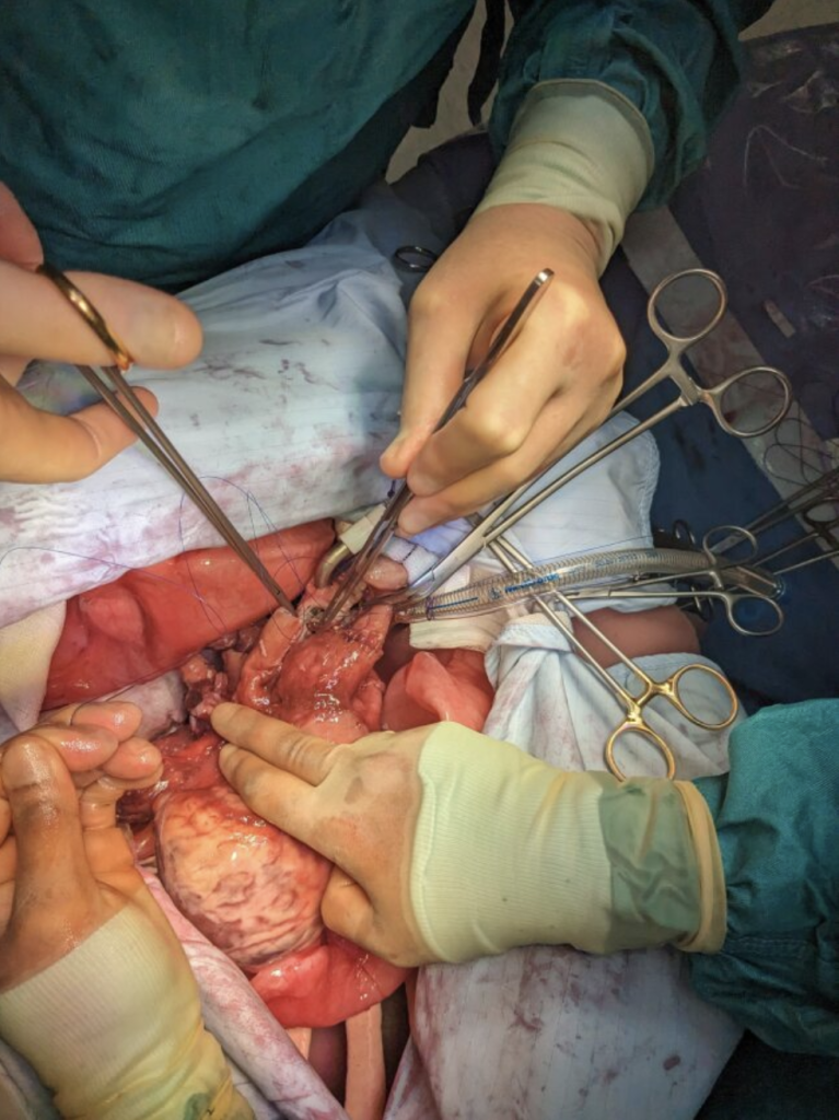 Surgeons practising organ transplant procedures with an NTU 3D printed heart model. Photo via Nottingham Trent University.
3D printed hearts enhance medical treatment
The NTU team are not the only researchers to identify the value of 3D printing for creating life-like organ models for medical training purposes.
Last year, a Massachusetts Institute of Technology (MIT)-driven project developed a process for 3D printing soft patient-specific robotic replicas of human hearts. Once fabricated, these models can accurately imitate the organ’s blood-pumping capabilities, helping clinicians to create personalized patient treatments.
The 3D printed hearts were developed using medical scans from 15 patients with aortic stenosis, a condition where the aortic valve narrows. Each scan was converted into a 3D model of the patients’ left ventricle (the primary pumping heart chamber) and aorta, which were then 3D printed using polymer-based ink. Once 3D printed, different valves could be implanted into the models to determine which design offers the best function and fit for the patient.
Elsewhere, researchers from the University of Minnesota created a functional 3D printed beating heart made from a specially developed, cell-laden biomaterial. The organ model, significantly smaller than a real human heart, was capable of pumping blood, offering the potential for drug testing and studying genetic diseases.
In 2020, researchers from Carnegie Mellon University developed what they claimed to be the world’s first full-size 3D printed heart models. The scientists employed a Freeform Reversible Embedding of Suspended Hydrogels (FRESH) technique, which involved the extrusion of eco-friendly alginate polymer into a custom-made gelatin container.
Through this approach, the team aimed to work with surgeons to 3D print patient-specific clinical models for surgical planning and pre-planning applications.
You might also like:
Breakthrough skin model accelerates development for testing and beyond: This achievement has implications for improving the testing of skin care products and potentially enhancing skin healing methods. Led by Associate Professor Paul Dalton from the Phil and Penny Knight Campus for Accelerating Scientific Impact at the UO, the research relies on Dalton’s novel 3D printing technique. Published in the journal Advanced Functional Materials, this technique enables the creation of a two-layered artificial skin, with each layer separated by a membrane, mirroring the structure of natural skin.
* This article is reprinted from 3D Printing Industry. If you are involved in infringement, please contact us to delete it.
Author: Alex Tyrer-Jones
Leave A Comment