Custom 3D printed models tailored to individual patients are helping surgeons remove oral cancers with greater precision, according to new research from The Ohio State University Comprehensive Cancer Center – Arthur G. James Cancer Hospital and Richard J. Solove Research Institute (OSUCCC – James). In 92 percent of head and neck surgeries that used a 3D model during the operation, complete tumor removal was achieved, compared to 74 percent of surgeries performed without one. Researchers report that this approach allows for more accurate excision of malignant tissue while preserving healthy structures, potentially reducing the need for follow-up treatment.
OSUCCC – James, a cancer research and treatment institution based in Columbus, Ohio, developed the technique in collaboration with Ohio State’s College of Engineering. The in-house Medical Modeling, Materials and Manufacturing Lab (M4 Lab) fabricates patient-specific anatomical models derived directly from imaging data. These models provide surgeons with physical guides that replicate the patient’s tumor boundaries and nearby structures before and during complex operations.
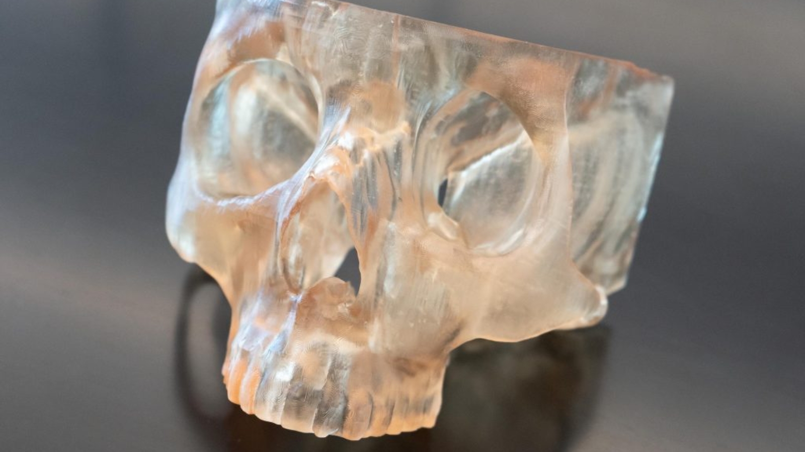
 3D printed patient-specific skull model produced at the M4 Lab. Photo via OSUCC – Arthur G. James Cancer Hospital/Richard J. Solove Research Institute
“The precision of what we take out is critical to ensure we get the whole tumor, but not so much that we’re devastating the patient’s function in the long term and taking out things that don’t need to be removed,” said Kyle VanKoevering, MD, an otolaryngologist at OSUCCC – James and medical director of the M4 Lab. “This 3D modeling being completely personalized to each patient is really helping improve the precision in the operating room.”
VanKoevering and his team compared outcomes for 68 patients with head and neck cancers that had invaded bone. Thirty-seven patients received 3D printed models for intraoperative use, while 31 underwent surgery without them. Nearly all participants were current or former tobacco users (94.6 percent). The group that used 3D models achieved better negative surgical margins, meaning the surrounding tissue showed no cancer presence, compared with those treated without visual aids.

 Dr. Kyle VanKoevering (right) and medical student Matthew Marquardt. Photo via OSUCC – Arthur G. James Cancer Hospital/Richard J. Solove Research Institute.
“This model is especially critical in cancers that have invaded bone, because tumor boundaries are often less visible or palpable,” VanKoevering said. “Our 3D models are built based on the patient’s actual tumor imaging, so it gives us a much better visual map at the patient’s bedside for removing the cancer as completely as possible while also sparing important structures and tissue to maintain function after surgery.”
The study, published in the September 2025 issue of Oral Oncology, is the first to evaluate how 3D modeling affects cancer control in surgery. Matthew Marquardt, the study’s corresponding author and a third-year medical student, led the project through the Pelotonia Scholars Program, which funds student-led cancer research. “This really sets the stage for larger studies looking at how 3D modeling can enhance surgery planning and precision, not just in the field of head and neck cancer surgery but in other areas that involve bone and soft tissue, like orthopedics,” Marquardt said. “Long term, our hope is that this work will enable other surgeons to use this technology across the country to help improve people’s lives and improve cancer outcomes.”
 The Ohio State University Comprehensive Cancer Center – Arthur G. James Cancer Hospital and Richard J. Solove Research Institute located in Columbus, OH. Photo via OSUCC – Arthur G. James Cancer Hospital/Richard J. Solove Research Institute.
The research team included Taylor Freeman, Amanda Pancake, Joseph Lee, MD, James Rocco, MD, PhD, Matthew Old, MD, Stephen Yang, MD, Lauren Miller, MD, Catherine Haring, MD, Nolan Seim, Enver Ozer, MD, Amit Agarwal, MD, Rachel Herster, Teri Snyder, and Megan Malara.
Manufacturing on Demand
The M4 Lab’s integration within OSUCCC – James enables surgeons to collaborate directly with engineers to convert imaging data into tangible surgical guides. This approach demonstrates how additive manufacturing is being applied to clinical oncology, linking digital modeling with hands-on surgical planning. As patient-specific fabrication becomes faster and more cost-effective, researchers anticipate broader adoption of similar in-house labs in academic hospitals.
For more information about cancer care and research at OSUCCC – James, visit cancer.osu.edu or call 1-800-293-5066.
You might also like:
3D Systems Secures FDA Clearance to Broaden Adolescent Use of VSP Orthopedics Platform: “This regulatory clearance removes a significant friction point for adoption in the pediatric/adolescent orthopedic oncology segment. Surgeons at leading centers have been using off-label or compassionate use solutions for years; this decision immediately converts those cases into routine clinical practice and opens the U.S. adolescent bone sarcoma and deformity market to our platform. We are thrilled to now offer these solutions to an expanded and underserved patient population,” said Ben Johnson, senior vice president of medical technology at 3D Systems.
* This article is reprinted from 3D Printing Industry. If you are involved in infringement, please contact us to delete it.
Author: Anyer Tenorio Lara
Leave A Comment