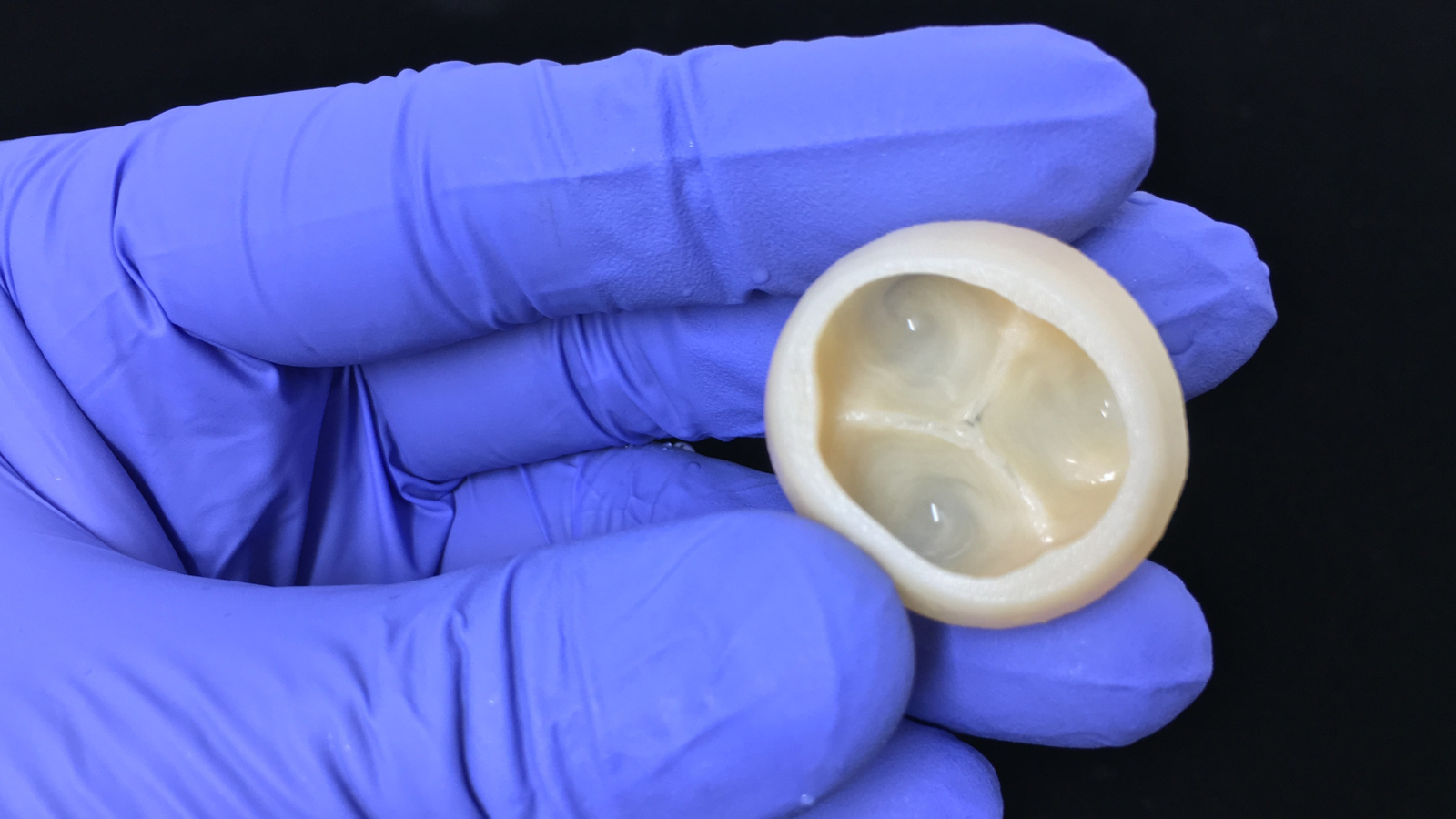
Scientists from Carnegie Mellon University (CMU), Pennsylvania, have used a novel 3D bioprinting method to build functional parts of the human heart.
According to a study published in Science, an advanced version of Freeform Reversible Embedding of Suspended Hydrogels (FRESH) technology was developed to 3D print collagen for small blood vessels, valves, and beating ventricles.
FRESH technology is patented to FluidForm, a Massachusetts-based medical startup. Professor Adam Feinberg, CTO and co-founder, FluidForm, and Principal Investigator at the Regenerative Biomaterials and Therapeutics Group, CMU, said:
“We now have the ability to build constructs that recapitulate key structural, mechanical, and biological properties of native tissues. There are still many challenges to overcome to get us to bioengineered 3D organs, but this research represents a major step forward.”

3D bioprinting soft materials
A common hurdle in 3D bioprinting is supporting complex structures made from soft materials and proteins, including collagean. FRESH uses a non-newtonian gel as a support material for such items. The properties of the gel allow a needle printhead to move through them as though they were liquid, overcoming the collapse or sagging of soft scaffolds.
Feinberg and CMU researchers applied rapid pH change during the FRESH process to drive self-assembly within a buffered support material. Mike Graffeo, CEO of FluidForm, adds, “The FRESH technique developed at CMU enables bioprinting researchers to achieve unprecedented structure, resolution, and fidelity, which will enable a quantum leap forward in the field. We are very excited to be making this technology available to researchers everywhere.”
Last year, Feinberg and his team developed an algorithm capable of selecting the optimal parameters for soft-material 3D printing, termed the Expert-Guided Optimization (EGO) method. This algorithm combines expert judgement with 3D printer optimization data to expedite new material development.
Manufacturing on Demand
3D bioprinted heart models
3D bioprinted heart models from human MRI data were created using CMU’s FRESH method as proof-of-concept showing the potential to build advanced scaffolds for a wide range of tissues and organ systems.
Following this, smaller cardiac ventricles 3D bioprinted with human cardiomyocytes, or cardiac muscle cells, were created to demonstrat a more solid structure, with wall-thickening up to 14%.
Nevertheless, the CMU team recognized the difficulties of generating the billions of cells required to 3D print larger tissues, as well as the undefined regulatory process for clinical translation of their method.
“3D bioprinting of collagen to rebuild components of the human heart” is co-authored by Andrew Lee, Andrew Hudson, D. J. Shiwarski, J. W. Tashman, T. J. Hinton, S. Yerneni, J. M. Bliley, P. G. Campbell, and Adam Feinberg.
* This article is reprinted from 3D Printing Industry. If you are involved in infringement, please contact us to delete it.
Author: Tia Vialva


Leave A Comment