Although 3D printing provides many users with the ability to create and innovate new designs and products, this technology can also be used to fill in the gaps. This can be outdated in terms of industrial components, real vehicles, submachine guns, or blood vessels that need to be recreated, parts of the human body need to be rebuilt and equipped with implants after such severe trauma, or the latest research shows that whether it is In law enforcement or in scientific institutions, anthropological findings and skeletal remains need to be reconstructed.
In the “Effective Method to Use Laser Surface Scanning for 3D Digital Reconstruction of Scattered Skeletal Remains,” authors from India and the United Kingdom discuss the remodeling, which may lose part of the bone 3D printing. In this study, they focused on reshaping part of the human skull and reconstructing the mandible. Historically, forensic anthropologists have superimposed photography and photography to identify missing persons, but due to areas of bone loss (for example, certain parts of the skull may be damaged), they have encountered challenges in the reconstruction process.
Today, the analysis of human remains relies on the following methods:
- 3D imaging
- X-rays
- Micro CTs
- Laser scanning
- Structured-light scanning
- 3D photogrammetry
Over the years, 3D digitization has provided clear advantages in many projects (usually in museums or research institutions), thereby making affordable copies for scientific communication, criminal forensics, teaching, museum displays, etc. The forensic department has benefited a lot worldwide and contributed to the work of other professionals.
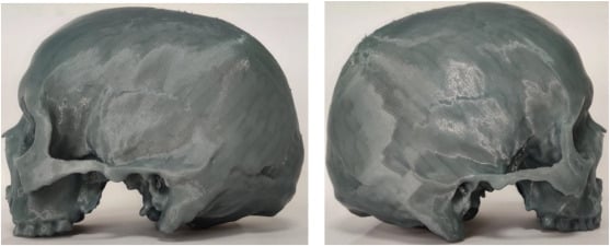
Researchers were able to use the NextEngine 3D laser scanner, Geomagic Studio 13 for surface reproduction and Flashforge Guider 2 3D printers to obtain two human skulls from the Skeleton Archives of the Gujarat University of Forensic Sciences for 3D scanning.
The skull model is printed with PLA 3D, and the size is 280×250×300 mm.
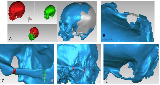
“A direct articulation was established between zygomatic process of maxilla and temporal bone after reconstruction of zygomatic arch,” stated the researchers. “The results were confluent with that mentioned in literature.”
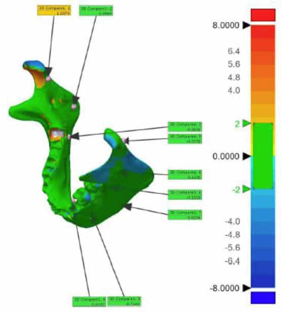
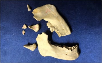
The mandible can also be obtained through Gujarat University of Forensic Sciences. The replica is made of vulcanized silicone and then filled with tartar. The researchers divided the replica into nine pieces.
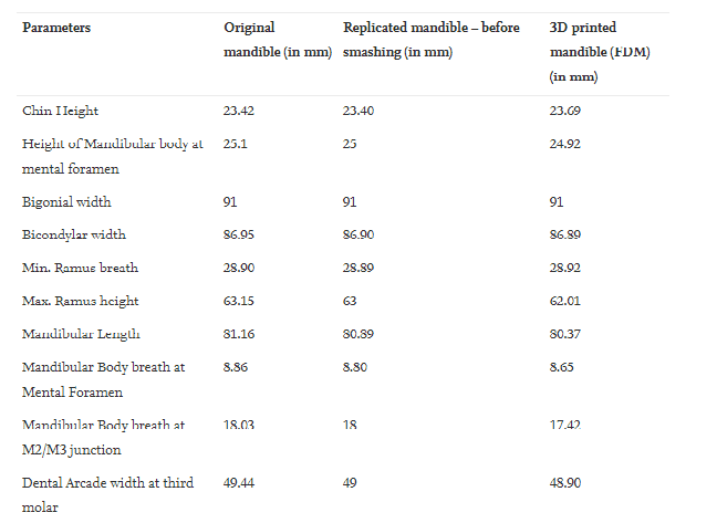
The obtained measurement results prove that the 3D printed model can be used for “various morphological analysis.”

Many different projects involving bone reconstruction rely on scanning a good side or complete area and then mirroring incomplete data. In this study, the incomplete bow was created using skull-like data. The researchers concluded that this model will be applicable to court as well as forensic anthropology and medicine.