Personalized precision medicine is on the rise. New tools and advanced technologies bring doctors closer to patients, and provide customized treatments and equipment to better serve each unique person.
In the advancement of medical 3D printing technology, a huge contribution has been made to the entire healthcare field. For patients, new tools and treatment methods developed through 3D printing can bring new comfort and personalization to treatment. For doctors, this newly available technology can give people a deeper understanding of complex cases, provide new tools, and ultimately improve the level of care.
From surgical planning models to 3D printed vasculature and bioreactors, read on to discover 3 ways 3D printing can get started in healthcare and why many medical professionals are excited about the potential of this technology in medicine .
Case 1 – 1:1 3D Model Constructed from CT Scan
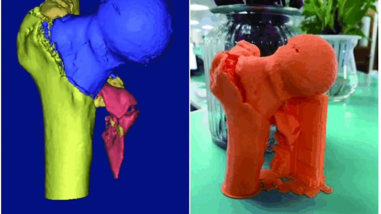
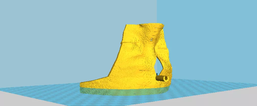
A few days ago, patient Huang accidentally slipped and felt severe pain in the right hip. Since then, he had difficulties in standing and moving. X-ray examination revealed a right femoral intertrochanter fracture.
The patient’s right femur had a deep fracture, surrounded by many important nerves and blood vessels. Cai Xiyu, deputy director of the Department of Traumatology and Joint Surgery, CUHK Hospital, used 3D printing technology to demonstrate the fracture of his right femur. The shape of the fracture, the number and location of the fracture were visually displayed in a three-dimensional manner, so as to make a more precise surgical plan.

According to doctor Cai Xiyu, in the past, orthopedic surgeries used X-rays or CT scans to examine the condition and make surgical plans. According to the patient’s CT reconstruction data, a 1:1 3D model was constructed, allowing doctors to understand the fracture condition more accurately. Then we can choose better surgical incisions, rehearse intraoperative steps, and tailor the best treatment plan for the patient.
Subsequently, the surgical team performed a closed reduction and intramedullary nail internal fixation (Intertan Surgery) for the comminuted fracture of the right intertrochanteric fracture to minimize the degree of invasiveness during the operation. At present, the patient has recovered smoothly and returned to work.
Case 2 – Surgical Planning with Full-scale 3D Printed Model
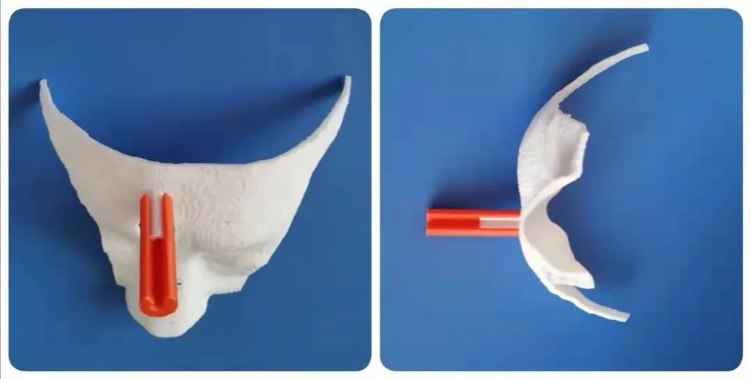
The 62-year-old patient Tao, suffers from long-term dialysis for uremia and has intermittent abdominal pain for nearly one month. The CT examination revealed an abdominal aortic aneurysm.
Aortic angiography confirmed that Tao’s abdominal aortic aneurysm was located in the visceral area of the abdominal aorta, and he also suffered from a penetrating ulcer of the abdominal aorta. The ulcer is adjacent to the superior mesenteric artery and the celiac artery and involves blood supply to the gastrointestinal tract, liver, kidney and other organs. The treatment is much more difficult than the general abdominal aortic aneurysm.
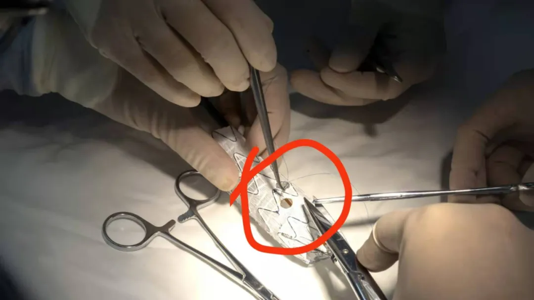
Doctor Wang Geng of the Department of Vascular Intervention in Zhongshan Hospital carefully assessed his condition and all aspects of the whole body. Considering that the patient has coronary heart disease, uremia and other diseases, if he chooses a surgical operation, the trauma will be too large.
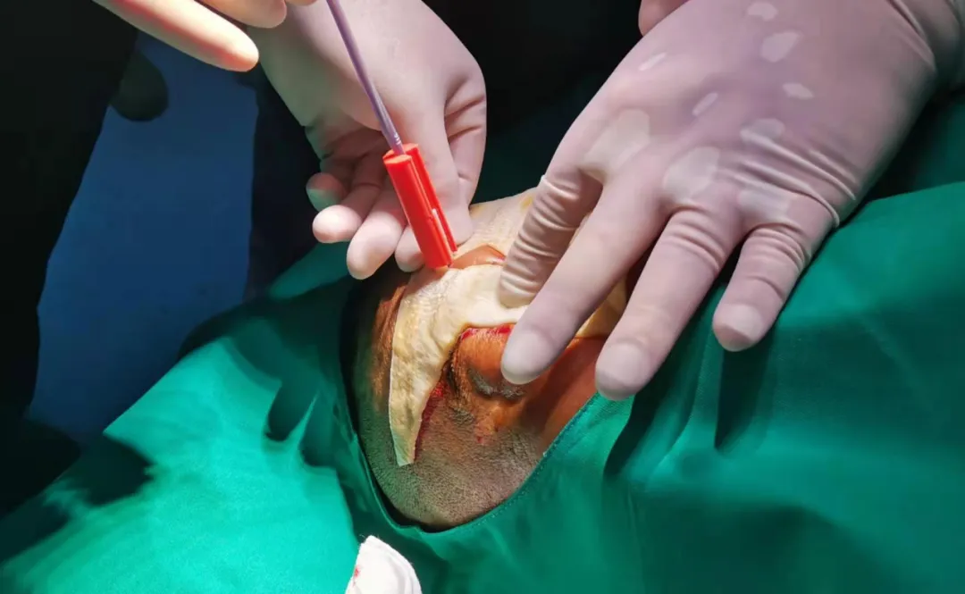
Director Wang used 3D printing technology to reconstruct the patient’s 1:1 abdominal aortic aneurysm model. The stent-graft was accurately opened outside the body and repeated exercises. During the operation, he led the team to pass the bilateral femoral artery and the right brachial artery through three puncture points. The important blood vessels on the artery are positioned, and the external “double window” technology is used on the stent to reserve the position of the superior mesenteric artery and the celiac artery. Then the stent is reassembled, and the superior mesenteric artery is used as the base point. The opening accurately connects the superior mesenteric artery and the celiac artery.
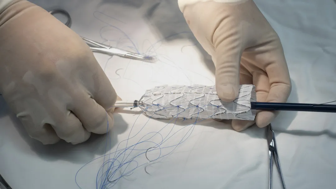
The difficulty of this treatment lies in the precise docking of the stent with the two vessels after the window is opened. Once any one of the vessels is sealed off in a dislocation, it can lead to organ loss and even life-threatening. The radiography showed that the patient’s abdominal aortic aneurysm was well isolated, and the blood flow of the superior mesenteric artery and the celiac artery was unobstructed. The patient recovered 7 days after the operation.
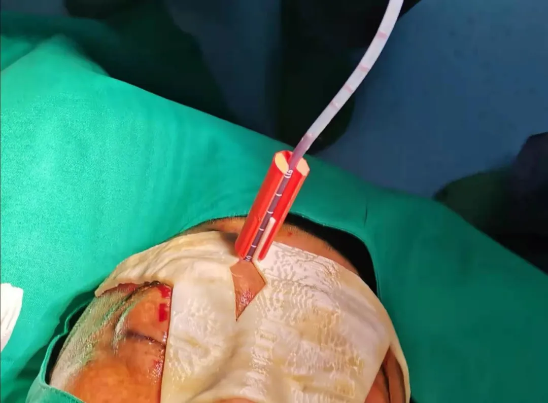
Case 3 – 3D Printed Surgical Guide for the Removal of High-density Hematoma
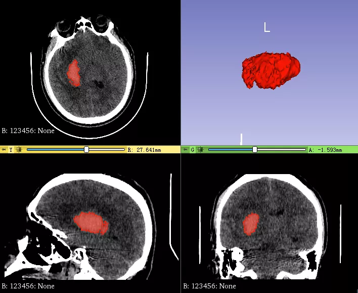
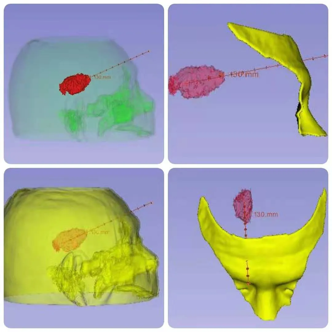
Gao, a 47-year-old male, was admitted to a hospital in Ningbo for non-surgical treatment with “headache after onset with left limb weakness” ten days ago. He has poor spirit, low muscle tone of the left upper and lower limbs, and grade 0 muscle strength. His bilateral eyes squint slightly to the right.
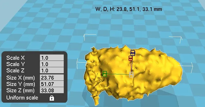
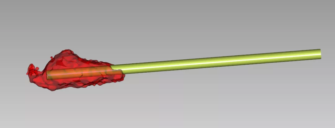
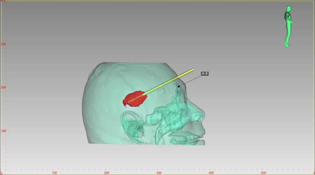
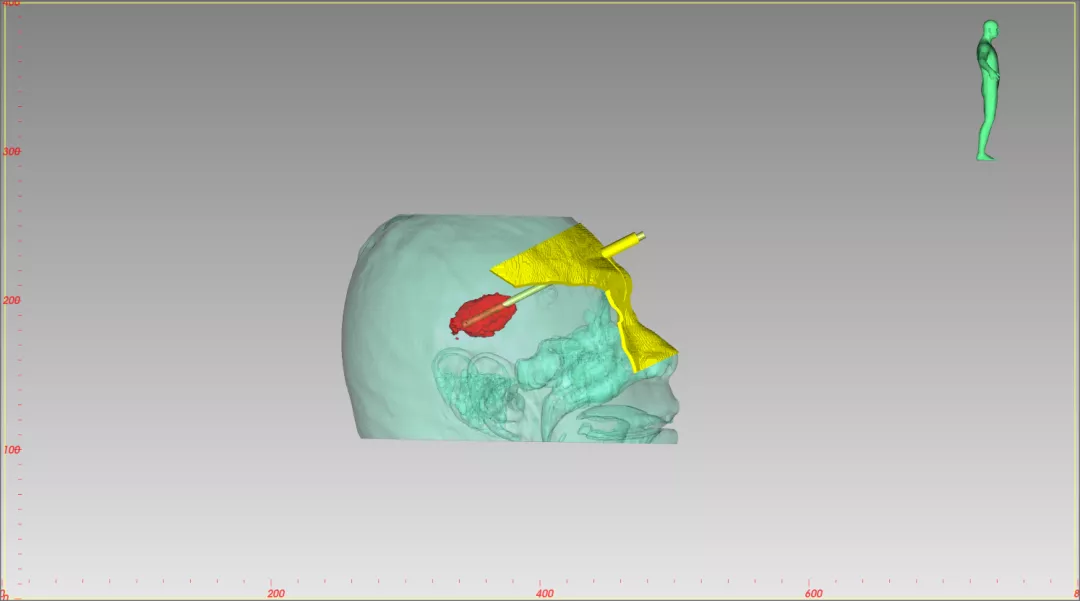
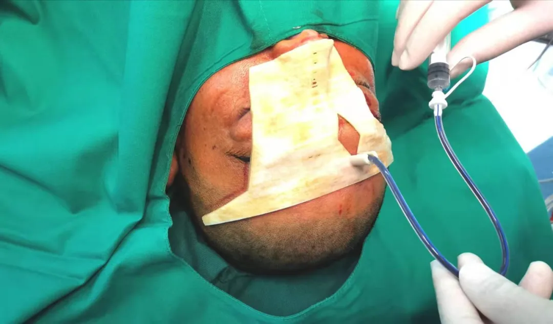
It was calculated that the high-density hematoma was 13.46ml after ten days, but considering the patient’s condition, continuing non-surgical treatment may lead to worse condition. In order to minimize surgical trauma and maximize the recovery of limb function, the doctor selected minimally invasive catheter drainage. Since the largest hematoma transverse diameter is 23.76mm, the long axis puncture slightly deviates, which may lead to puncture failure or deviation, the doctor decided to apply 3D printing technology for precise positioning and puncture.
15 hours after the operation, the drainage tube drained about 20ml of a dark brown hematoma. The patient reported that the headache was relieved. Physical examination: left-hand muscle strength 2-3, left upper limb 1-2, left lower limb muscle strength 1-2 level.
The image and data of hematoma
Surgical planning with 3D Printing


Leave A Comment
You must be logged in to post a comment