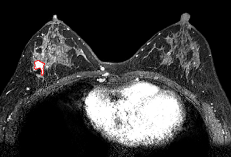
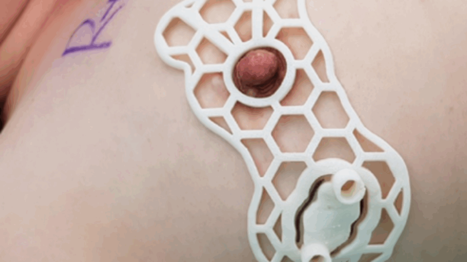
Researchers from the Korea-based Asan Medical Center have 3D printed surgical guides that could help cancer patients to retain more of their breasts after surgery.
The scientists found during testing that they were not only able to customize their devices to each patient, but they could save tissue up to 1cm from the tumor. In future, the team aim to further develop their technique in order to enable tailor-made guides to become more widely-adopted within breast cancer procedures.
“Hospitals have to use MRI to determine the scope of surgery, however, [current] methods cannot directly mark the extent of the cancer on the breast,” said Kim Nam-gook, who led the research. “The research is significant in that the accuracy of the team’s 3D printing surgery guide has been proven even in early breast cancer.”

Adopting additive to treat cancer patients
Ductal Carcinoma In Situ (DCIS) is the earliest form of breast cancer, and although it’s not considered imminently life threatening, treatment is often required to stop it from spreading. Thanks to an increase in active screening examinations, a growing number of DCIS cases are being discovered, but many women still require Breast-Conserving Surgery (BCS).
Hospitals actively try to save as much breast tissue as possible during BCS by only removing the affected areas, but precision remains key in optimizing the operation. Despite this, in up to 58 percent of cases, conventional scans underestimate the size of the tumor, preventing surgeons from achieving the best patient outcomes.
Wire-Guided Localization (WGL) is the most commonly used method of improving accuracy, but it risks cutting the affected tissue, or even losing the wire. Alternatively, a team from the University of South Florida has developed a Radioactive Seed Localization (RSL) approach, however its adverse radiation effects have prevented its wider adoption.
According to the Korean researchers, the WGL and RSL techniques are also flawed because they are not compatible with the more accurate MRI scans. Consequently, the team has opted to develop an MRI-guided technique that could enable the creation of devices that are optimized to fit each patient individually.

3D printing and testing the team’s guides
In order to test their theory, the scientists gained the consent of eleven breast cancer patients, to use the additive guide during their procedures. Once they’d got approval, the team conducted MRI scans on their volunteers, and separated the cancerous tissues from the healthy ones using Materialise’s Medical software.
The scientists modelled their guide with a 0.5 cm margin of error to ensure the patient’s safety. To optimize the performance of their devices, the team then designed it with a hole for attaching it to the nipple, and a suprasternal notch, column and grooves for marking the tissue and its underlying internals.
The guides were then attached to the patients, before a blue dye was injected through the device’s columns, to indicate the tissue being removed. Given that all eleven patients continued their treatment after surgery, the team later opted to measure their success using the accuracy of their products instead.
Manufacturing on Demand
Overall, the team’s MRI-based approach proved to be more accurate than existing methods, yielding a median margin of error of 11mm. Additionally, in three of the study’s cases, the tumor was not seen on ultrasonography but it was observed via MRI scans, which prevented the need for invasive biopsies to be performed.
Although the team conceded that their sample size was small, they viewed their 3D printed guide to be a success. In fact, due to the variances observed in the conditions of their patients, the scientists believe that their device has proven the ability to improve accuracy, regardless of cancer’s stage of development.
3D printing’s cancer-fighting remedies
Researchers have utilized 3D printing technologies in a myriad of ways in recent years, in order to better combat cancer in each of its various forms.
Researchers from Washington State University (WSU) have created a soy-infused 3D printed scaffold that’s capable of fighting off cancer cells. Once mixed into bioprinted tissues, the team believes that their bone-like compound could actively improve the post-operative care of young cancer patients.
Similarly, a Spanish group of scientists have developed a hydrogel that’s capable of mimicking the behaviors of human lymph nodes. By combining PEG with heparin, the researchers were able to 3D print a structure that allowed T-cells to proliferate and combat cancer cells more effectively.
Elsewhere, researchers from Virginia Commonwealth University have 3D printed a live model of tumor cells, to better understand how the disease progresses. AM enabled the team to more effectively mimic the key steps of cancer dissemination, and the model could eventually allow them to test anti-cancer drugs.
The researchers’ findings are detailed in their paper titled “.”
The study was co-authored by Zhen-Yu Wu, Aisha Alzuhair, Heejeong Kim, Jong Won Lee, Il Yong Chung, Jisun Kim, Sae Byul Lee, Byung Ho Son, Gyungyub Gong, Hak Hee Kim, Joon Beom Seo, Sei Hyun Ahn, Namkug Kim and BeomSeok Ko.
* This article is reprinted from 3D Printing Industry. If you are involved in infringement, please contact us to delete it.
Author: Paul Hanaphy


Leave A Comment