Researchers from J. Stefan Institute and the University of Ljubljana in Slovenia have achieved a scientific first by 3D printing tiny functional structures directly inside living cells.
Using a high-precision laser technique called two-photon polymerization (2PP), they created custom polymer microstructures including microlasers, tracking barcodes, and even a 10 µm model of an elephant, all within the cytoplasm of living HeLa cells.
Published in arXiv, the approach allows these structures to form in place without harming the cell’s internal environment, offering a new way to engineer cells from the inside out. The study was led by Matjaž Humar, Physicist at J. Stefan Institute and University of Ljubljana, who conceived and supervised the project.
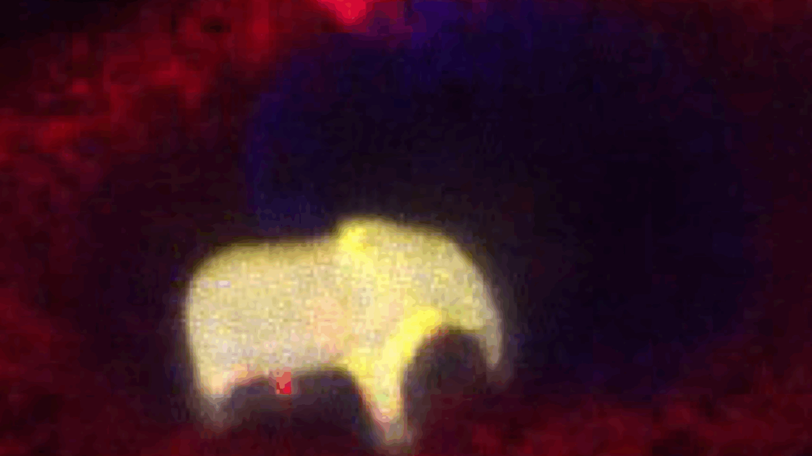
 Confocal image of a 10 µm elephant printed inside a live HeLa cell. Image via J. Stefan Institute.
Direct fabrication inside living cells
The process begins with the injection of a photoresist droplet into the cell, followed by laser scanning to trigger polymerization only at the point of focus. As the laser moves through the droplet in a programmed pattern, it creates a solid 3D structure while the remaining material dissolves naturally. This method allows precise, in-situ fabrication of microdevices without introducing external particles.
The researchers used the Photonic Professional GT2 system from Nanoscribe for printing, which employs a femtosecond 780 nm laser suited for submicron resolution. For materials, they tested several photoresists from the same manufacturer and selected IP-S for its low toxicity and water solubility. A 10 µm droplet typically dissolves within a few hours, giving a two-hour window to complete printing. Its compatibility in both liquid and solid forms was critical to maintaining cellular viability throughout the process.
To understand how the procedure affected cell health, the team ran a 24-hour viability study. Of the cells with printed structures, 55 % remained alive. Cells injected with inert silicone oil showed 44 % viability, and those receiving only buffer reached 50 %. Even untouched controls saw 10 % cell death, likely due to extended handling. These figures suggest membrane penetration during injection was the main source of stress, not the structures themselves.
In some cases, cells continued normal activity, including one that successfully divided and passed the printed structure to a daughter cell. Confocal imaging confirmed that internal components, including the nucleus, adapted around the structure without compromising integrity, demonstrating that cells could accommodate these foreign elements without immediate disruption.
To assess potential distortion caused by the curved droplet, the team used simulations and microscopy. Laser focus shift was limited to 0.5 µm, and over 90 % of the droplet volume maintained resolution finer than 400 nm. Scanning electron microscopy confirmed structural fidelity, with printed features as small as 260 nm, approaching the resolution of bulk printing.
The researchers demonstrated how these prints could be used in real-world applications by creating 3D barcodes composed of stacked 4 × 4 grids, enabling 61 bits of data per structure, more than enough to uniquely tag every cell in the human body. These predesigned codes allow consistent labeling and long-term tracking in complex biological studies.
They also printed diffraction gratings inside cells, which produced distinct light-scattering patterns when illuminated. Because the diffraction profile depends on structure geometry and orientation, these prints allowed remote optical readout of cell position or rotation. The team then produced microlasers by printing high-refractive-index, dye-doped photoresist droplets. These 9 µm resonators emitted laser light when excited but were significantly more toxic, with 80 % of cells dying or visibly stressed within 22 hours.
Manufacturing on Demand
Though current limitations include material toxicity and cell loss from injection, the approach opens promising directions. Structures could be designed to interact mechanically with organelles, alter cell shape, or isolate specific regions of the cytoplasm. Researchers could probe biochemical pathways, simulate disease conditions, or introduce tools for sensing and signal processing entirely from within the cell.
This technique avoids the limitations of particle ingestion, which only works for certain cell types and often traps material inside vesicles. By building directly in the cytoplasm, scientists gain access to a broader range of cells and greater control over where and how synthetic structures interact with native biology.

 Viability of cells with printed structures. Image via J. Stefan Institute.
2PP’s growing role in biomedicine
Two-photon polymerization is increasingly becoming a go-to method for fabricating high-resolution microstructures in the medical field.
High-resolution micro-scale 3D printing company UpNano GmbH developed UpFlow, a photopolymer designed for 2PP, enabling fast and precise fabrication of micro-environments for embryo culture.
Working with Australian IVF specialist Fertilis, the material was used with the NanoOne 3D printer to produce a 3D printed device featuring 0.05 mm structures that combined fertilization, embryo culture, and cryopreservation in one unit. This streamlined approach reduced the need for manual embryo handling and cut production time from two weeks to just four hours. The system also lowered implantation cycles by 30-40 %, while ensuring optical clarity and efficient post-processing of fine microchannels.
Elsewhere, Aston University integrated the Quantum X bio 3D printer to advance its biomedical research capabilities through 2PP. Designed for high-precision fabrication, the system allowed researchers to replicate complex cell orientations found in organs like the brain and liver.
It supported studies on astrocyte behavior, the blood-brain barrier, soft liver tissue models, and drug delivery mechanisms. Housed in a specially controlled environment, the printer marked the first UK installation of its kind and formed part of broader efforts in tissue engineering and drug discovery across multiple research departments.
You might also like:
Irish researchers reveal new electrically active implant for spinal repair: Researchers from the Royal College of Surgeons in Ireland (RCSI) University of Medicine and Health Sciences and Trinity College Dublin have developed a 3D printed implant that may support the repair of spinal cord injuries by delivering electrical stimulation to damaged nerve tissue.
* This article is reprinted from 3D Printing Industry. If you are involved in infringement, please contact us to delete it.
Author: Ada Shaikhnag
Leave A Comment