A team at The Prince Charles Hospital in Brisbane has carried out a complex and high-stakes procedure to replace the majority of a man’s aorta after scans revealed it had swollen to an abnormal size.
To prepare for the operation, surgeons turned to a full-scale 3D printed model of the patient’s aorta to better visualize the complexity of his case. According to ABC News, the man is in his late fifties from Queensland, and was unaware his aorta had gradually expanded to nearly 8 cm, which vascular surgeon Samantha Peden explained is far beyond the normal range of 2 to 3.
He had previously undergone surgery in 2017 to repair a tear in the artery, but the vessel continued to dilate over time. Doctors suspect an underlying connective tissue disorder, though no specific diagnosis has yet been confirmed. As the aortic wall thinned and the risk of rupture increased, it became clear that replacement surgery was the only option to prevent a potentially fatal outcome.
“You can never tell with certainty when it was going to rupture, but it definitely was at the end of its ability to expand,” said Dr. Peden.
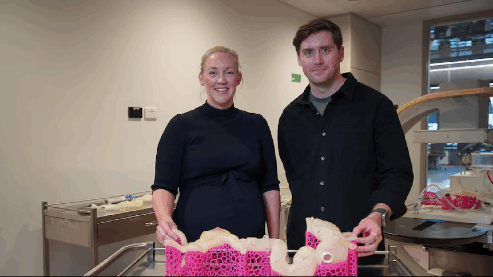
 Vascular surgeon Dr. Samantha Peden and industrial designer Liam Georgeson with a 3D model of a patient’s stretched aorta. Photo via ABC News.
A second chance at life
Rather than rely solely on standard imaging, the team collaborated with the Herston Biofabrication Institute (HBI) to produce a highly detailed 3D model of the affected artery. Engineers and designers used digital scans of the patient’s body to build a replica using Stratasys J750 Digital Anatomy 3D printer.
The process took four days, with the printer layering materials of varying textures to mimic both the structure and the tactile feel of human tissue. Having access to this physical model gave surgeons a clearer understanding of the patient’s unique anatomy and allowed them to plan with greater precision than conventional two-dimensional scans would allow.
The operation lasted nine hours and brought together vascular and cardiac surgeons in a tightly coordinated effort. To safely access and replace the upper section of the aorta, the medical team induced circulatory arrest. The patient’s body was cooled, and his heart temporarily stopped to allow blood flow to halt entirely for 20 minutes.
But these minutes came with a lot of risks. “[Patients] can wake up with a stroke or they might not wake up,” Dr. Peden said, with possibilities of Paraplegia, kidney and liver failure. Yet within the critical window, surgeons removed the compromised section and replaced it with a synthetic graft made of a flexible and waterproof fabric capable of withstanding the intense pressure of blood circulation.
Almost three weeks after the operation, the patient remains in hospital but has moved out of intensive care. According to his doctors, “he’s recovering really well” showing no major complications and full movement in his arms and legs. He is expected to begin intensive rehabilitation shortly, with a follow-up surgery planned later this year to replace the lower portion of the aorta.
The team at The Prince Charles Hospital completes an average of just six full aortic replacements each year due to the complexity involved. Surgeons likened the process to precision plumbing, requiring exact reconnections between major vessels under tightly controlled conditions.
Manufacturing on Demand
Looking ahead, the HBI is continuing its work on creating patient-specific models that simulate blood flow using pump systems. These dynamic replicas are designed to help surgeons rehearse procedures under conditions that more closely mirror the real physiological environment.

 The life-size model was used by surgeons to better plan life-saving surgery. Photo via Queensland Health.
Patients specific models for enhanced surgical planning
Patient-specific 3D printed models play a key role in surgical planning by giving surgeons a clear, hands-on view of complex anatomy. They help with everything from pre-op planning to team coordination, making procedures smoother and more precise.
In 2023, 3D printer OEM Stratasys and multinational electronics firm Ricoh partnered to deliver on-demand, patient-specific 3D printed anatomical models designed to support surgical planning, diagnosis, and education. The service combined Stratasys’ Digital Anatomy printing technology with Axial3D’s Segmentation-as-a-Service solution and Ricoh’s certified manufacturing capabilities to produce lifelike models that closely replicated the texture and structure of real tissue.
Clinicians could securely upload medical scans to a cloud-based system, which generated 3D printable files. These were then printed at Ricoh’s ISO 13485-certified facility and shipped directly to hospitals, reducing turnaround times from weeks to days and removing the need for in-house printing expertise or infrastructure.
One year before this, researchers from the Mayo Clinic, Georgia Tech, and the Medical University of South Carolina developed a 3D printed spinal model as a proof of concept to address gaps in surgical training.
Their goal was to highlight the limitations of traditional training methods, which can be costly, difficult to access, and incompatible with neuromonitoring systems used during surgery. To provide a more practical and affordable alternative, they created the lateral access training model (LATM), featuring anatomically accurate 3D printed lumbar vertebrae designed to support realistic, hands-on learning for spinal procedures.
You might also like:
Tessella Biosciences develops new bioink to 3D print realistic lung tissue: Canada-based McMaster University-spinout Tessella Biosciences has developed a new bioink that allows scientists to 3D print soft lung tissue capable of expanding and contracting like real lungs.
* This article is reprinted from 3D Printing Industry. If you are involved in infringement, please contact us to delete it.
Author: Ada Shaikhnag
Leave A Comment