
An Israeli anesthesia team recently used 3D printing and virtual reality technology to create an accurate model of the respiratory tract of a 7-year-old girl as part of the surgery to remove her lungs.
With cancer of the bone and soft tissue that has spread to her lungs, the young girl needs to remove part of her right lung. In order to properly prepare for the surgery, her doctors at the Surasky Medical Center in Tel Aviv used her virtual reality procedures for airways and lungs as well as accurate 3D printed plastic models so that they could perform the surgery ahead of time.
“Despite the extensive adult experience, our familiarity with one-lung ventilation (OLV) in the very young pediatric population is limited,” explains Dr. Ruth Shaylor of the Division of Anesthesia, Pain and Intensive Care, Tel Aviv Sourasky Medical Center, the lead author on the report.
“We used a combination of 3D printing and virtual reality bronchoscopy to develop a personalized airway plan, thereby reducing the possibility of repeated trials due to airway manipulation during surgery.”
3D printing personalized airway plan
The disease that troubled the young girl spread to her lungs and is called Ewing’s sarcoma, which is a rare type of cancer that affects the bone or the tissues around it.
When preparing to operate on the patient, the doctor’s main consideration was her short stature, because she was only 18 kg during the operation. The use of conventional catheters and equipment commonly used in lung surgery in elderly patients may cause potential complications.
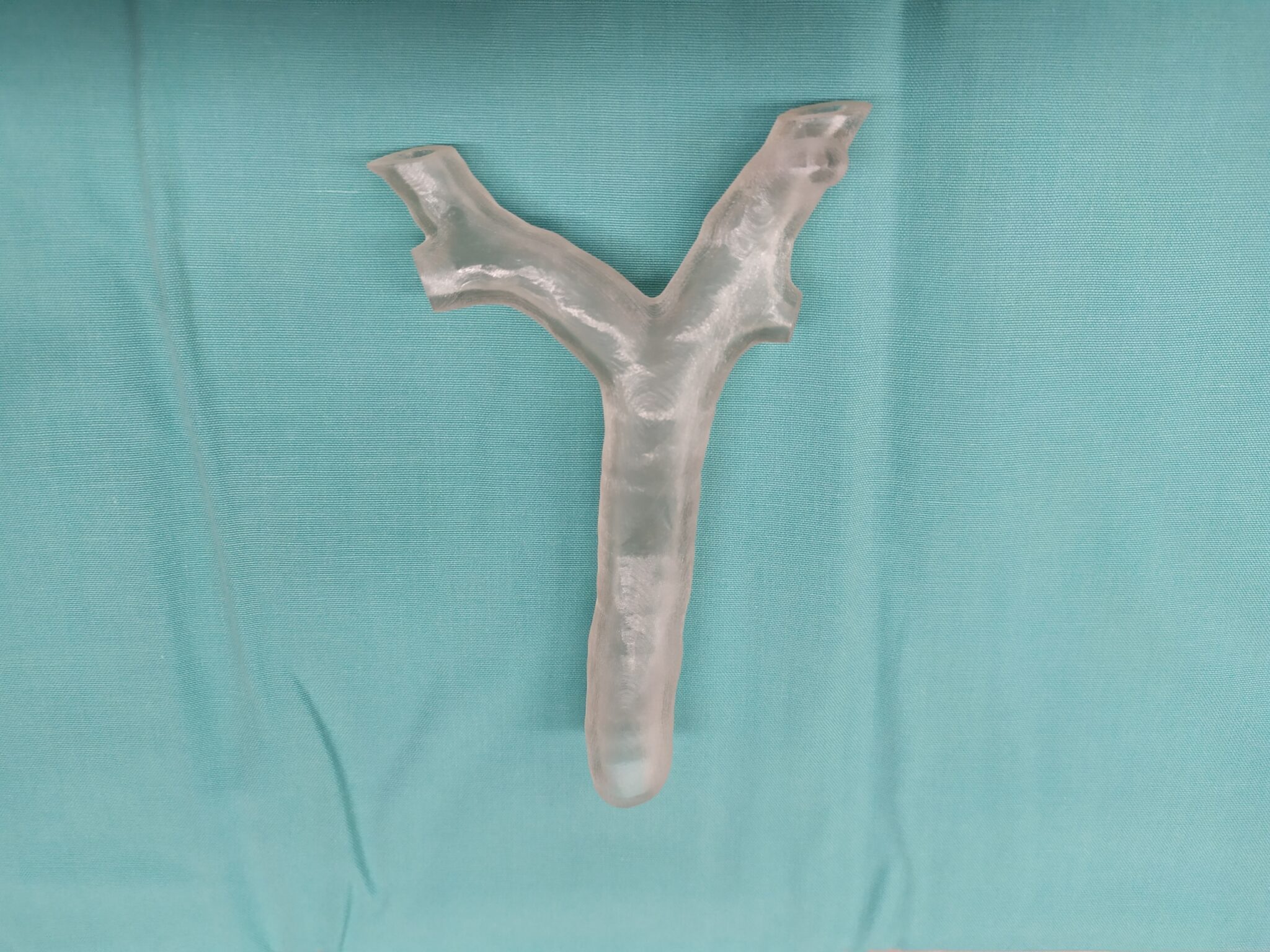
Therefore, the doctor chose to use a CT scan to create a virtual reality program for her airway/air duct and lungs. The day before the operation, the attending anesthesiologist spent some time on the virtual reality simulator to familiarize him with the patient’s airway anatomy.
After that, the model was 3D printed and used to develop a personalized airway plan. The 3D model is made of hard plastic, because anything softer may be inaccurate, which means that something larger than the patient can bear can be used.
VR and 3D printed models mean that some equipment originally planned for this operation has been changed. Once the girl is anesthetized, they can safely isolate and ventilate the left lung and undergo surgery to remove part of the right lung. “During the operation, the attending anesthesiologist reported that there was a good correlation between the patient’s anatomy and the VR model, and the operation could be performed as planned,” Dr. Shaylor commented. “At the end of the operation, the catheter and equipment can be easily pulled out and the patient is restored to stability.”
Dr. Shaylor continued that he explained that to date, 3D printing in anesthesia has mainly been used for educational purposes, but it has the potential to further assist surgery: “3D printing in anesthesia was not obtained in the preoperative plan for specific patients Make the most of it. The same is true of the use of virtual reality in virtual anesthesia. We have successfully combined these two technologies to develop a personalized airway plan for pediatric patients. The final airway plan is very different from the plan made using standard imaging techniques The same. This reduces the number of lung isolation attempts that would have been performed on the patient.”

Medical services for patients
Many occasions and plans have been established in which 3D printing is used for patient-specific preoperative plans to help surgeons perform their procedures.
For example, the Puget Sound Healthcare System of Veterans Affairs (VA) announced in 2019 a two-year collaboration with the University of Washington School of Medicine (UW) to create a 3D printed model for patients to help treat the mitral valve disease. Abnormal heart.
GE Healthcare and Formlabs also announced a collaboration aimed at making it easier for clinicians to 3D print patient-specific anatomical models from imaging data.
The 3D printed, patient-specific medical equipment has also been used outside of the preoperative plan and has been used in the procedure itself. In early 2020, the 3D printed patient-specific airway stent developed by doctors at the Cleveland Clinic received FDA approval to be implanted in patients.