Doctors at the University of Toledo Medical Center are exploring a new way to treat chronic wounds, using the patient’s own fat.
For this approach, physicians begin by taking high-resolution images of the affected area which are processed with artificial intelligence (AI) to generate a 3D model of the patient’s wound. From there, a small sample of fat is removed, typically from the patient’s abdomen. That tissue is then processed into a printable form and loaded into the Aplicor 3D, a bioprinter developed by biotechnology research company Tides Medical. The result is a graft shaped specifically to fit the wound.
“It’s almost like printing a puzzle piece,” said Dr. Munier Nazzal, who leads the Division of Vascular, Endovascular Surgery and Wound Care. “We’re able to make a graft that is an exact fit for the wound, and that graft is going to promote healing. It’s not magic — it’s not healing tomorrow, but it’s healing faster than we see from our traditional methods of treatment. We’re very early on in this, but it has shown good results.”
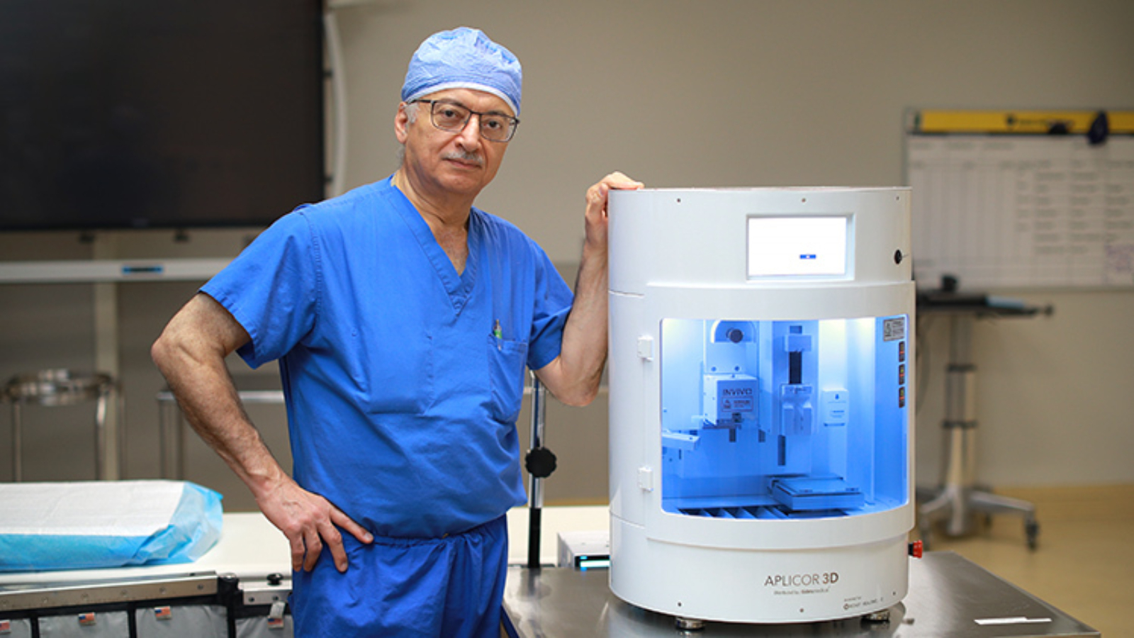
 Dr. Munier Nazzal, a UToledo Health vascular surgeon, poses with a 3D graft printer. UToledo Health is one of the first healthcare systems in the country to treat chronic wounds using 3D printed grafts derived from a patient’s own fat cells. Photo via UToledo.
Printed grafts support healing from within
Unlike traditional skin grafts, the printed material is not a replacement for lost tissue. It does not close the wound through coverage alone. Instead, it creates an environment that encourages the body to repair itself. The printed graft sits in place, shielding the injury while stimulating the natural healing process from within.
For patients living with long-term, non-healing wounds, this offers a different kind of hope. Often linked to diabetes, poor circulation, or autoimmune conditions, these injuries do more than cause discomfort.
They can lead to serious infections, hospital stays, and in severe cases, amputations. Existing treatments like cleaning the wound, improving blood flow, and using oxygen therapy can be effective, but for many patients the progress is slow or nonexistent.
That was the case for Jeffrey Paul, one of the first patients at UToledo Health to try the new procedure. Diagnosed with an autoimmune blistering disorder, Paul had spent months dealing with a persistent lesion on his ankle.
Despite regular visits and consistent care, the wound refused to heal. When his care team suggested trying the new 3D printed graft, he agreed. Within two weeks, he noticed something he hadn’t seen in a long time: the wound was finally closing.
The technology behind the procedure arrived at UToledo Health with the help of a former patient who donated funds for the printer. That same contribution is now being used to expand the hospital’s wound care capabilities.
Two additional tools are being added to the program, including one that can detect high levels of bacteria in a wound and another that measures oxygen levels in the surrounding tissue.
Manufacturing on Demand
“It’s our responsibility to stay up to date on the latest technology and knowledge so we can best serve our patients. These new technologies help us tailor the treatment plans to the specific patient needs,” Nazzal said. “We are so grateful for our donor’s faith in our program and their generosity that has allowed us to add these cutting-edge tools.”
Advances in bioprinted skin continue to grow
Bioprinted skin has been drawing increasing attention as researchers investigate its potential uses in medicine. These lab-grown skin models offer a platform for studying various diseases, testing new therapies, and providing alternatives to animal testing in scientific research.
In line with this, Pusan National University researchers developed a 3D bioprinting method to create functional fat tissue that supports wound healing and skin regeneration. Using a hybrid bioink made from 1% adipose-derived extracellular matrix and 0.5% alginate, the team provided an environment that promotes fat cell maturation and stability.

 The conditions for 3D bioprinting of adipose tissues involved the use of adipose-derived decellularized extracellular matrix and alginate as a hybrid ink. Image via PNU.
To ensure proper cell function, printed fat units were kept under 600 μm in size and within 1000 μm of each other to support paracrine signaling. In mouse models, the bioprinted tissue accelerated healing, improved vascularization, and influenced proteins critical to skin cell migration and remodeling.
Three years ago, researchers from the University of Birmingham and the University of Huddersfield developed a bioprinting technique called Suspended Layer Additive Manufacturing (SLAM) to produce a tri-layered skin equivalent for treating chronic wounds. The method allowed hypodermis, dermis, and epidermis cells to be printed in a gel-based support structure, preserving their arrangement until the gel was removed.
The resulting skin model accurately simulated both the chemical and mechanical properties of natural skin. In early tests using pig skin, the printed patch showed signs of tissue integration and wound repair after two weeks of culturing, indicating promising potential for human applications.
You might also like:
Bioengineered islets retain shape and function with 90% viability in 3D printing breakthrough: Researchers at Wake Forest University and University of Miami have developed a way to 3D print insulin-producing human islets using a bioink made from alginate and decellularized pancreatic tissue. The work was presented at the ESOT Congress 2025.
* This article is reprinted from 3D Printing Industry. If you are involved in infringement, please contact us to delete it.
Author: Ada Shaikhnag
Leave A Comment