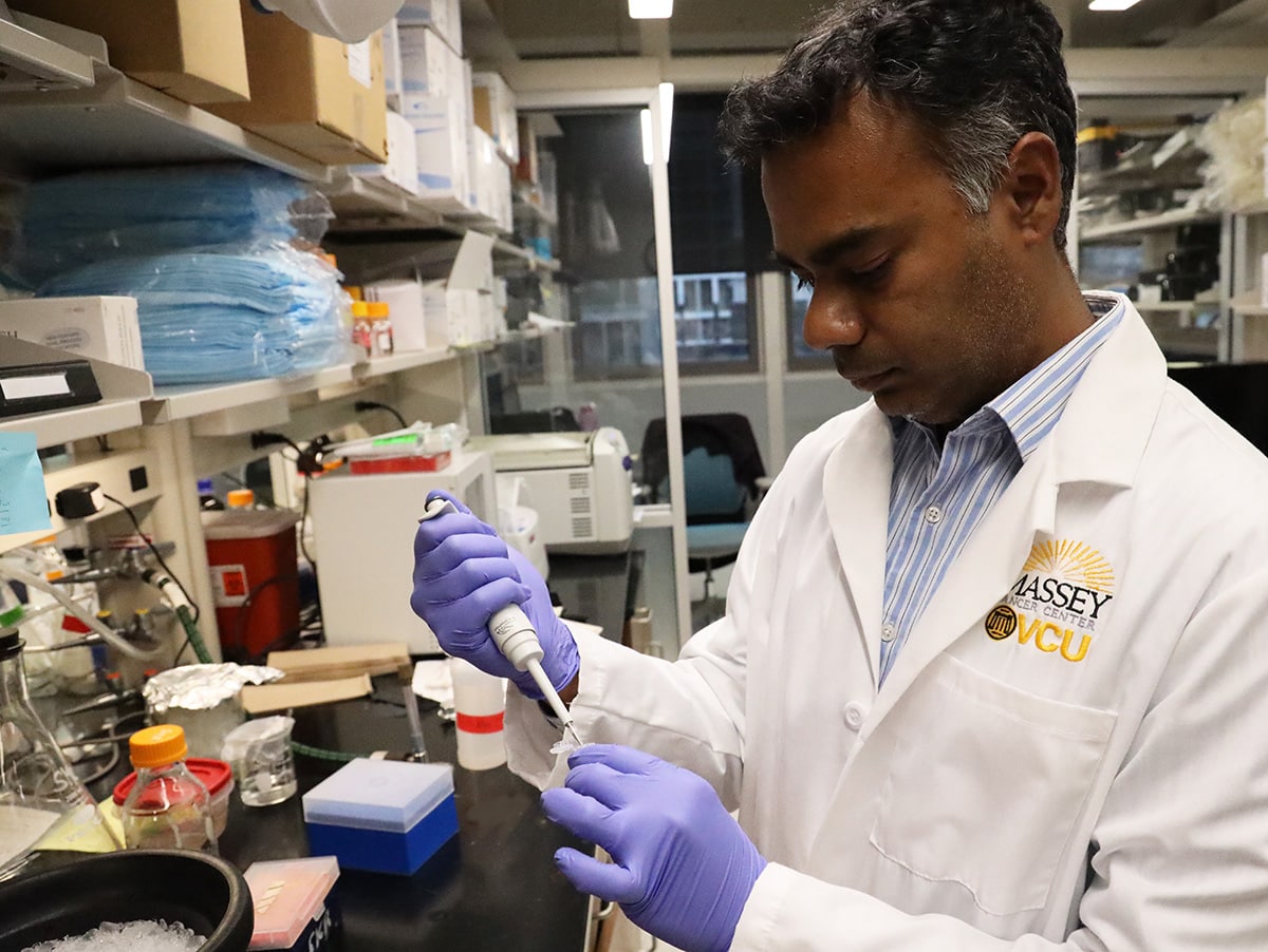
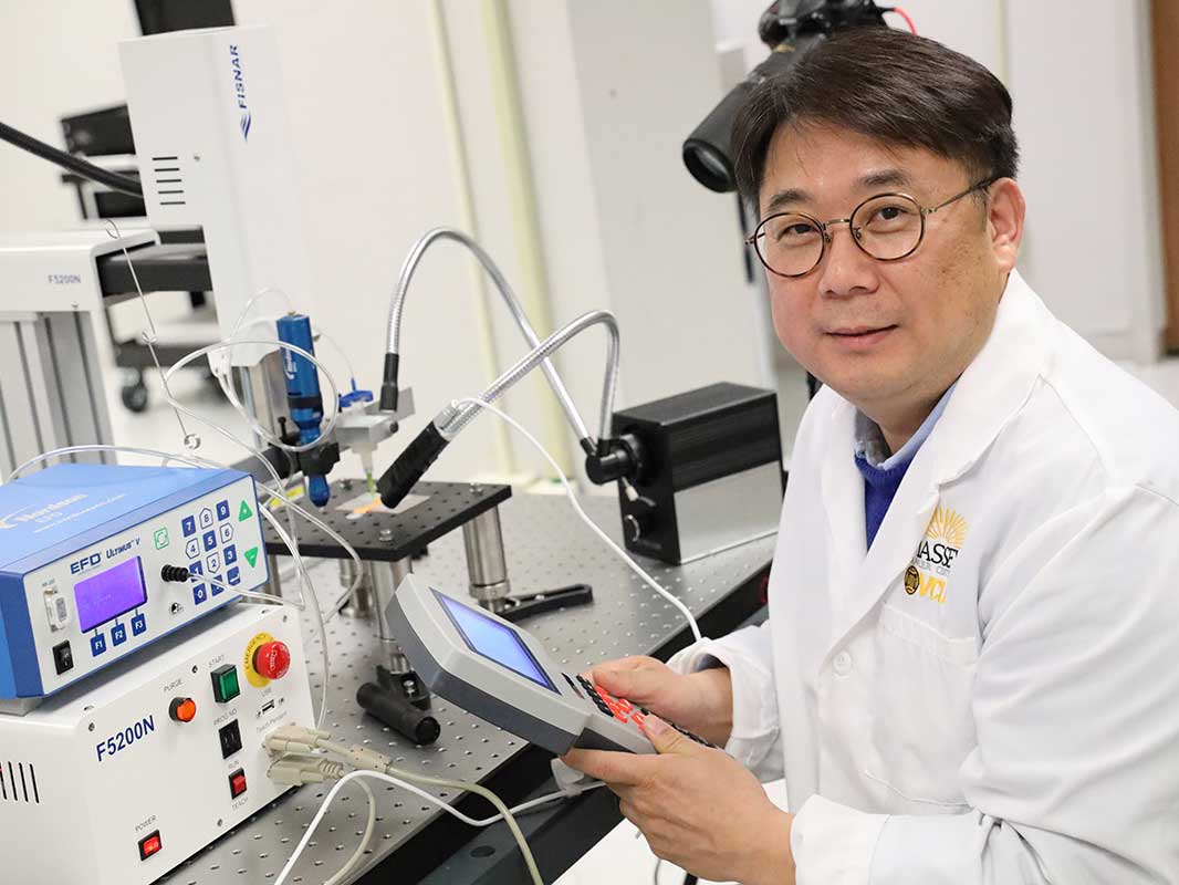
The assistant professor at the Virginia Commonwealth University School of Humanities and Sciences has used 3D printing to create real-time models of tumor cells, which can give cancer researchers a better understanding of disease progression.
Assistant Professor Daeha Joung worked with a team of researchers led by Dr. Michael McAlpine of the University of Minnesota (UMN) to invent new methods for constructing tumor constructs. This new technology can accurately locate and control living cells, thereby more effectively simulating the key steps of cancer spread. Now at VCU, Joung is using this method to enable researchers at the University ’s Massey Cancer Center to maximize the growth of cancer cells for research and to evaluate the effectiveness of anti-cancer immunotoxins.
The evolution from 2D to 3D printing in cancer research
Researchers from the German European Molecular Biology Laboratory discovered in December 2018 that it is difficult to accurately replicate the characteristics of the natural tumor microenvironment in a two-dimensional structure. It was found that 3D cell culture can establish cell-cell interactions to better simulate the specificity of real tissues, proceed at a faster rate, and exhibit more complex behaviors.
However, according to UMN researchers, cancer metastasis remains the main prognosis and therapeutic challenge of the current model because the movement of tumor cells is regulated by multiple chemical signals. Therefore, there is a need for a more accurate model that should combine these elements of the tumor microenvironment and be able to study the multi-level interactions that occur at a distance from the human body. Joung explained: “Many strategies have been developed to overcome these obstacles, but they have not been completely successful.” “I can use physics, nanotechnology, and engineering techniques to overcome these obstacles and provide unique insights that can be useful for cancer. New treatment options open the door. ”
The team’s new approach addresses these limitations by using 3D bioprinting and origami-inspired self-folding techniques to create complex multi-material, multi-scale, and multi-functional 3D structures. These allow precise spatial control to integrate multiple combinations of living cells and supporting matrices, enabling tissue engineering for promising biomedical applications.

Manufacturing of 3D printed cell control capsules
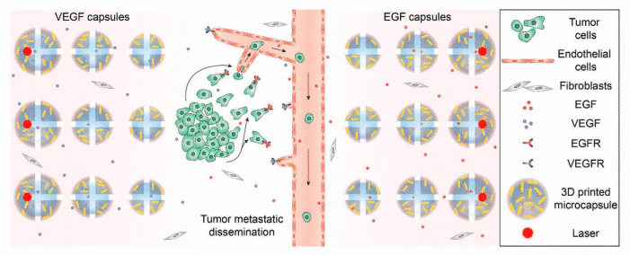
3D printed stimuli-responsive capsules allow researchers to control the cells inside these structures. The microcapsules contain water cores, effective molecular functional factors, and plasmon gold nanorods (AuNRs), which can trigger the release of chemical cues in the 3D matrix. Once activated using laser radiation, the research team was able to manipulate cell behavior at a local level. When used in conjunction with 3D cell printing, it is possible to create complex tumor constructs controlled using multiple chemical signals.
To demonstrate the use of capsules for spatial control, the researchers printed a series of cores containing different volumes, which can be precisely triggered by deploying ultraviolet lasers. Using 3D printing, researchers can test five sets of migration samples in parallel under the same conditions. This method also allows him to add materials when necessary, and customize the resulting cells to show different behaviors.
As a result of this technology, Massey University scientists will be able to provide Joung with cancer tissue samples and then specify the specific designs or tumor structures they need to manufacture. Joung will then use robotic mechanisms and customized 3D printing technology to adjust the number and specific configuration of the unit types to be copied and printed.
In turn, this could enable future researchers to screen new anti-cancer drugs and test patient-specific diagnosis and treatment strategies. Joung said: “From an engineering perspective, cancer is cancer, so my laboratory can produce many different types of tumor tissue mimics.” “If someone is interested in lung cancer, we can print lung cancer cells. If someone is Breast cancer cells are of interest and we can also print them out, “he added.
Joung ’s ambition is to design an integrated platform for an advanced 3D system for use in cancer nerve regeneration models, thereby providing new opportunities for testing treatments to treat diseases.

3D printing method to fight cancer
Scientists and researchers in other academic institutions have also used 3D printing to create real-time 3D cell models with the goal of better understanding and treating cancerous tumors.
In April 2020, researchers from the United States and Germany proposed a new method to study glioblastoma (GBM), an aggressive brain cancer. They used human brain cells and biological materials with perfusion vascular channels for long-term culture and drug delivery. 3D imaging technology has been broadly used for non-invasive assessment of tissue structure.
A master student from the University of Waikato in New Zealand announced in August 2018 that she plans to use real cells to grow and test cancer tumors based on the design of a 3D printed plastic tumor model. The basis of these bioprinting models is a series of hand-webbed hemispheres similar to breast cancer tumors.
Researchers at the Kathholieke Universiteit Leuven in Belgium and the Indian Institute of Technology in Hyderabad, India released a study in June 2017 to study how to use 3D printing for cancer research. Although both papers emphasize the potential of using 3D printing to create a microenvironment, Indian researchers have proposed the possibility of accurately placing cells and replicating the tumor microenvironment in vivo.
The researchers’ findings were detailed in a paper titled “3D Bioprinting In Vitro Metastasis Model by Reconstructing the Tumor Microenvironment”, which was published in the journal Advanced Materials by Carolyn M. Meyer, Daeha Joung, Daniel A co-authored Vallera, Michael C. McAlpine and Angela Panoskaltsis-Mortari.

